Oncological radiology leverages advanced medical imaging technologies like X-rays, CT scans, MRI, PET, and ultrasound to visualize tumors, aiding cancer detection and diagnosis by providing detailed cross-sectional and metabolic information. These tools empower radiologists to stage diseases accurately, plan personalized treatments, and monitor therapy response, ultimately improving patient outcomes.
In the realm of cancer care, early detection and precise diagnosis are pivotal. Oncological radiology, a specialized field within medical imaging, plays a crucial role in this process. This article delves into the diverse landscape of oncological radiology, exploring common imaging techniques like X-rays, CT scans, and MRIs used for cancer detection. It also highlights advanced scanning technologies that further refine diagnosis. Additionally, it discusses the art of interpreting these images to guide effective treatment strategies.
Understanding Oncological Radiology: Unveiling Visual Diagnostics
Oncological radiology, a specialized field within medical imaging, plays a pivotal role in cancer detection and diagnosis. This branch focuses on using various imaging techniques to visualize abnormalities within the body, particularly those associated with tumors. By employing advanced technologies such as X-rays, computed tomography (CT), magnetic resonance imaging (MRI), positron emission tomography (PET), and ultrasound, radiologists can uncover subtle changes indicative of cancerous growths.
The power of oncological radiology lies in its ability to provide detailed, high-resolution images that offer crucial insights into the nature and extent of cancers. For instance, CT scans excel at identifying solid tumors, while MRI offers unparalleled contrast to visualize soft tissue abnormalities. PET scans, on the other hand, are adept at detecting metabolic activity associated with cancer cells, enabling early detection even before visible tumors emerge. This comprehensive array of imaging tools equips healthcare professionals with vital information for informed decision-making in cancer management.
Common Imaging Techniques for Cancer Detection
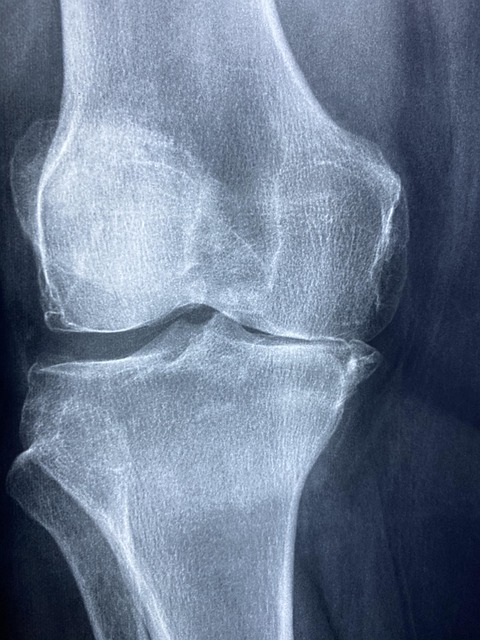
Cancer detection and diagnosis have significantly advanced with the integration of various medical imaging techniques, particularly in oncological radiology. Common methods include X-rays, which are often the first line of examination for initial screening and assessing bone fractures or lesions. Computed Tomography (CT) scans offer a more detailed view, generating cross-sectional images that can identify tumors, their size, and location. This non-invasive technique is invaluable for oncologists when planning treatment strategies.
Magnetic Resonance Imaging (MRI) is another powerful tool, creating intricate images of soft tissues and organs, aiding in the detection of cancer spread or response to therapy. Positron Emission Tomography (PET) scans are used to visualize metabolic activity, helping identify cancerous cells and assessing treatment effectiveness. Additionally, Ultrasound imaging, though primarily used during pregnancy, can also detect tumors and guide biopsies, making it a versatile tool in oncological radiology.
Advanced Scans: Enhancing Diagnosis Precision
Advanced scans, often employed in oncological radiology, have revolutionized cancer detection and diagnosis. Technologies such as magnetic resonance imaging (MRI), computed tomography (CT) scans, and positron emission tomography (PET) offer unprecedented detail about a patient’s condition. MRI provides clear anatomical images, aiding in the identification of tumors and their spread. CT scans, with their high-resolution cross-sectional images, assist in detecting early signs of cancer, including small lesions that might be missed by other methods. PET scans, on the other hand, use radioactive tracers to highlight areas of abnormal metabolic activity, often indicating the presence of cancerous cells.
These advanced imaging techniques not only help in identifying the location and size of tumors but also assess their functional characteristics, such as blood flow and metabolism. This comprehensive view enables radiologists to make more accurate diagnoses, determine the extent of disease (staging), and plan personalized treatment strategies for each patient.
Interpreting Images: Pathways to Effective Treatment
Interpreting medical images is a critical component of cancer detection and diagnosis, especially in oncological radiology. Radiologists use their expertise to analyze various imaging modalities such as X-rays, CT scans, MRIs, and PET scans, which provide valuable insights into the body’s internal structures. By examining these images, healthcare professionals can identify suspicious growths, tumors, or abnormalities that may indicate the presence of cancer.
Accurate interpretation allows for early detection, enabling oncologists to devise effective treatment plans. This process involves meticulous attention to detail, as even subtle changes in tissue structure or pattern can be indicative of cancer progression or response to therapy. Advanced imaging techniques and artificial intelligence tools further enhance the precision and speed of diagnosis, ultimately leading to better patient outcomes.
Oncological radiology plays a pivotal role in cancer detection and diagnosis, offering diverse imaging techniques that range from conventional X-rays to advanced scans like MRI and PET. By leveraging these technologies, radiologists can accurately identify tumors, assess their extent, and guide treatment strategies. Interpreting medical images requires specialized knowledge and expertise, ensuring patients receive effective care based on precise diagnostics. Understanding oncological radiology empowers healthcare professionals to navigate the complex landscape of cancer detection, ultimately enhancing patient outcomes.