Advanced imaging techniques like MRI and CT scans have revolutionized tumor imaging, enabling early detection and accurate characterization of brain tumors through high-resolution insights into anatomy. MRI uses magnetic fields and radio waves, while CT employs X-rays to generate detailed images. Future prospects include diffusion-weighted MRI, functional MRI, AI, and machine learning for improved diagnostic accuracy and personalized treatment plans in tumor imaging.
Brain tumors can be challenging to detect, but advanced imaging techniques are revolutionizing diagnosis. This article explores innovative methods for identifying brain tumors, focusing on Magnetic Resonance Imaging (MRI) and Computed Tomography Scans (CT). We delve into the strengths and applications of each technology, highlighting their role in improving patient outcomes. Furthermore, we discuss emerging trends in tumor imaging, offering a glimpse into the future where early detection and precision medicine will shape brain cancer care.
Advanced Imaging Techniques for Tumor Detection
Advanced imaging techniques have revolutionized tumor imaging, providing doctors with detailed insights into brain anatomy and pathology. Technologies such as magnetic resonance imaging (MRI) and computed tomography (CT) scans offer high-resolution images that can detect tumors at an early stage, when treatment options are often more effective.
These non-invasive methods utilize different physical principles to create visual representations of the brain. MRI employs magnetic fields and radio waves to generate detailed anatomical pictures, while CT uses X-rays to produce cross-sectional images. By analyzing these images for abnormalities, healthcare professionals can accurately identify the presence, size, location, and type of tumors, guiding personalized treatment plans.
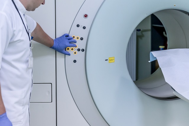
Magnetic Resonance Imaging: A Powerful Tool
Magnetic Resonance Imaging (MRI) stands as a powerful tool in the realm of tumor imaging, offering detailed and non-invasive insights into brain anatomy. This advanced technique leverages strong magnetic fields and radio waves to generate high-resolution images, enabling healthcare professionals to detect, diagnose, and monitor brain tumors with remarkable accuracy. MRI scans can distinguish between healthy tissue and abnormal growths, providing crucial information about the size, location, and characteristics of a tumor.
The versatility of MRI in tumor imaging is further enhanced by its ability to employ various contrast agents and specialized sequences. These innovations permit the detection of subtle changes within the brain, improving diagnostic precision. Moreover, MRI’s non-ionizing nature makes it a safer alternative compared to other imaging methods, making it an indispensable asset in modern neurology and oncology practices.
Computed Tomography Scans and Brain Tumors
Computed Tomography (CT) scans play a significant role in detecting brain tumors due to their ability to create detailed cross-sectional images of the brain. This non-invasive technique uses X-rays and advanced computer processing to generate high-resolution pictures, allowing healthcare professionals to identify abnormalities such as tumors, bleeding, or inflammation. CT scans are particularly valuable for quick assessments, pre-surgery planning, and monitoring treatment progress.
By utilizing contrast dyes, CT imaging can highlight suspicious areas in the brain, making it easier to distinguish between healthy tissue and tumor masses. Advanced CT scanning technologies, like high-resolution 3D imaging, enable more precise tumor characterization, enhancing the accuracy of diagnosis and treatment decisions. These scans are a crucial step in the early detection and management of brain tumors through effective tumor imaging strategies.
Future Directions in Tumor Imaging Technologies
The future of tumor imaging holds immense potential for early detection and precise characterization of brain tumors. Researchers are continually exploring innovative techniques to enhance diagnostic capabilities. One promising area is the development of advanced magnetic resonance imaging (MRI) sequences, such as diffusion-weighted MRI and functional MRI, which can provide detailed information about tumor biology and microstructure. These technologies enable the visualization of subtle changes in brain tissue, potentially allowing for earlier identification of malignant growths.
Additionally, the integration of artificial intelligence (AI) and machine learning algorithms is revolutionizing tumor imaging. AI models can analyze vast amounts of medical imaging data, learn patterns associated with specific tumor types, and assist radiologists in making more accurate diagnoses. This technology has the potential to improve inter-rater reliability, reduce false positives and negatives, and ultimately streamline the diagnostic process for brain tumors. Continued research and refinement of these cutting-edge imaging techniques will undoubtedly contribute to improved patient outcomes and enhanced understanding of neurological diseases.
Advanced imaging techniques, such as Magnetic Resonance Imaging (MRI) and Computed Tomography (CT) scans, have revolutionized tumor detection, providing invaluable insights into brain tumors’ complex nature. As technology advances, future developments in tumor imaging will undoubtedly enhance diagnostic capabilities further, leading to improved patient outcomes and a deeper understanding of neurological conditions. These cutting-edge tools are transforming the landscape of medical diagnosis and treatment, offering hope for more effective management of brain tumors.