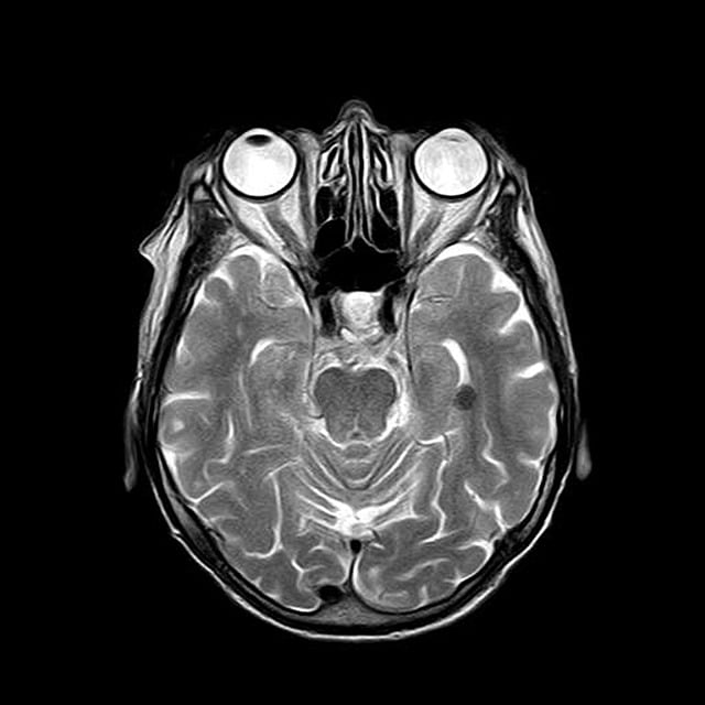
Soft tissue tumors present diagnostic challenges due to their diverse nature. Magnetic Resonance Imaging (MRI) with contrast agents offers a powerful tool for visualizing these tumors, differentiating benign and malignant lesions, and guiding treatment strategies through molecular imaging capabilities. By tracking biomarkers and metabolic activities, MRI enhances diagnosis accuracy, enables early-stage tumor detection, and supports personalized cancer care.
Magnetic Resonance Imaging (MRI) has emerged as a powerful tool in the early detection and diagnosis of soft tissue tumors. This non-invasive technique offers detailed insights into tumor characteristics, aiding oncologists in making precise treatment decisions. In this comprehensive guide, we explore the role of MRI in molecular imaging for cancer detection, delving into contrast agents, advanced visualization techniques, and how these innovations enhance diagnostic accuracy in identifying and understanding soft tissue tumors.
Understanding Soft Tissue Tumors: A Comprehensive Overview
Soft tissue tumors are a diverse group of growths that originate in the connective tissues of the body, including muscles, fats, ligaments, and blood vessels. Unlike bone or brain tumors, which have distinct structural characteristics, soft tissue tumors can be more challenging to diagnose and categorize due to their varied nature and location. These tumors often present as lumps or masses, but their appearance and behavior can vary widely depending on their type and where they form.
Molecular imaging for cancer, particularly using Magnetic Resonance Imaging (MRI), offers a powerful tool for understanding and detecting these subtle abnormalities. MRI provides detailed images of soft tissues, allowing healthcare professionals to differentiate between benign and malignant tumors based on their structural and molecular features. By analyzing the unique chemical composition and blood flow patterns within these masses, MRI enables doctors to make more accurate diagnoses and plan effective treatment strategies, ultimately improving patient outcomes in the management of soft tissue tumors.
The Role of MRI in Molecular Imaging for Cancer Detection
Magnetic Resonance Imaging (MRI) has emerged as a powerful tool in molecular imaging for cancer detection, offering unprecedented insights into the complex microenvironment of tumors. By utilizing specialized contrast agents and advanced scanning techniques, MRI enables the visualization of biological processes at a molecular level. This capability is particularly valuable in identifying and characterizing soft tissue tumors, which often present challenges for traditional diagnostic methods.
Molecular imaging through MRI allows healthcare professionals to track specific biomarkers associated with cancer cells, providing a more precise and targeted approach to detection. This non-invasive technique can detect early-stage tumors, monitor treatment response, and even predict patient outcomes. The detailed anatomic information provided by MRI, combined with its molecular sensitivity, makes it an invaluable asset in the battle against cancer, contributing to improved diagnostic accuracy and personalized treatment strategies.
Unlocking Tumor Characteristics: Contrast Agents and MRI
Magnetic Resonance Imaging (MRI) stands out as a powerful tool in the field of medical imaging, offering a unique window into the complex world of soft tissue tumors. One of its most significant advantages lies in its ability to utilize contrast agents, enhancing the visibility and characterization of tumors. These agents, composed of molecules that interact with magnetic fields, play a pivotal role in molecular imaging for cancer. By strategically injecting these substances into the patient, MRI scanners can distinguish between healthy tissues and abnormal growths based on their distinct signal intensities.
Through this enhanced contrast, healthcare professionals gain invaluable insights into tumor characteristics such as size, shape, location, and vascularity. Contrast agents, particularly those designed for molecular imaging, can even provide information about metabolic processes within the tumor, allowing doctors to make more precise diagnoses and tailor treatment plans accordingly. This advanced approach promises improved detection rates and enhanced therapeutic outcomes in the ongoing battle against cancer.
Advanced Visualization Techniques: Enhancing Diagnostic Accuracy
Advanced visualization techniques, such as molecular imaging for cancer, play a pivotal role in enhancing diagnostic accuracy with MRI. By tracking specific biological markers and metabolic activities within the body, these techniques offer a more detailed and nuanced view of soft tissue tumors. This allows radiologists to distinguish between benign and malignant lesions, identify tumor boundaries, and assess the extent of disease spread.
Molecular imaging for cancer leverages specialized agents that target specific molecular pathways or receptors expressed by cancer cells. These agents emit signals detected by MRI scanners, creating highly resolved images that highlight tumor biology and microstructure. This level of detail not only aids in precise diagnosis but also guides targeted therapies, improving patient outcomes and potentially reducing the need for invasive procedures.
Magnetic Resonance Imaging (MRI) plays a pivotal role in detecting soft tissue tumors through advanced molecular imaging techniques. By utilizing contrast agents, MRI provides detailed insights into tumor characteristics, enabling healthcare professionals to make more accurate diagnoses and develop tailored treatment plans. These innovative visualization methods have significantly enhanced the early detection and management of cancer, making MRI an indispensable tool in modern oncology.
