Biopsy-guided imaging is a revolutionary tool in cancer diagnosis and treatment, integrating histopathological analysis with advanced real-time imaging to accurately identify tumor types, grades, and molecular alterations through small tissue samples from specific regions of interest. This enhances surgical interventions and radiation therapy planning, enabling personalized medicine and improved patient outcomes by providing detailed insights into each tumor's unique properties, especially in addressing tumor heterogeneity. It combines biopsy data with functional imaging techniques for targeted therapies, achieving early cancer detection and safe, non-invasive tumor monitoring.
“Explore the cutting-edge world of functional imaging techniques, transforming tumor analysis and diagnosis. From biopsy-guided imaging, which enhances accuracy by combining invasive biopsies with advanced visualization, to non-invasive monitoring methods, these innovations offer comprehensive insights into cancer’s complexities.
Learn how functional imaging reveals tumor heterogeneity, enabling more precise treatment planning. Discover the latest advancements that promise improved patient outcomes, offering a glimpse into the future of oncology diagnostics.”
Biopsy-Guided Imaging: Enhancing Tumor Diagnosis
Biopsy-guided imaging plays a pivotal role in enhancing tumor diagnosis by combining the precision of histopathological analysis with the spatial context provided by advanced imaging technologies. This approach allows doctors to obtain small tissue samples from specific regions of interest within a tumor, ensuring accurate identification of cancer types, grades, and potential molecular alterations. By integrating this information into the corresponding anatomical images, healthcare professionals gain invaluable insights into the tumor’s characteristics and behavior.
Moreover, biopsy-guided imaging facilitates targeted treatment planning. Radiologists can pinpoint the location of suspicious lesions or areas of interest, enabling more effective surgical interventions or precise delivery of radiation therapy. This level of detail not only improves diagnostic accuracy but also contributes to personalized medicine by tailoring treatments to the unique properties of each tumor, ultimately leading to better patient outcomes.
Functional Techniques for Comprehensive Analysis
Functional imaging techniques play a pivotal role in comprehensive tumor analysis, providing valuable insights beyond what traditional anatomical imaging can offer. These advanced methods enable researchers and clinicians to study the biological activity and metabolic processes within tumors, leading to more accurate diagnoses and personalized treatment strategies. One such powerful tool is biopsy-guided imaging, which combines real-time visualization with histopathological correlation.
By integrating functional imaging with biopsy procedures, healthcare professionals can obtain dynamic data on tumor characteristics, such as glucose metabolism, blood flow, and oxygenation levels. This comprehensive analysis allows for a more nuanced understanding of the tumor’s microenvironment, identifying distinct regions within a single lesion that may exhibit varied behaviors. Consequently, these techniques foster more effective treatment planning, ensuring targeted therapies reach the most active areas of a tumor while minimizing damage to surrounding healthy tissues.
Navigating Tumor Heterogeneity: Visual Insights
Tumor heterogeneity poses a significant challenge in cancer diagnosis and treatment, as tumors often consist of diverse cellular populations with varying genetic profiles and metabolic activities. Functional imaging techniques offer powerful tools to navigate this complexity by providing visual insights into the tumor’s inner workings. By integrating biopsy-guided imaging approaches, healthcare professionals can gain a deeper understanding of tumor architecture, identifying distinct regions with unique characteristics.
This visual exploration enables the differentiation between tumor borders, core areas, and potential zones of cellular interaction or angiogenesis. The ability to visualize these nuances is crucial for making informed decisions regarding treatment planning. For instance, functional imaging can highlight areas of high metabolic activity or vascularization, guiding targeted therapies while minimizing damage to surrounding healthy tissues.
Advancements in Non-Invasive Tumor Monitoring
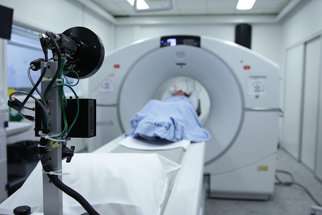
The field of medical imaging has witnessed significant advancements in non-invasive tumor monitoring, revolutionizing the way we detect and track cancerous growths. One notable innovation is biopsy-guided imaging, which combines real-time imaging capabilities with the precision of biopsy procedures. This technique enables doctors to take tissue samples from suspicious lesions without invasive surgery, providing a less risky alternative for patients.
By utilizing advanced imaging modalities like magnetic resonance imaging (MRI) and computed tomography (CT), healthcare professionals can now visualize tumors more accurately and obtain biopsy samples with higher success rates. Biopsy-guided imaging plays a crucial role in early cancer detection, helping to diagnose and stage tumors at earlier stages when treatment outcomes are generally better. This non-invasive approach not only enhances patient safety but also opens doors for more efficient and effective cancer management strategies.
Functional imaging techniques have revolutionized tumor analysis, offering non-invasive methods like biopsy-guided imaging that enhance diagnosis and provide comprehensive insights into tumor heterogeneity. These advancements enable more precise treatment planning and monitoring, ultimately improving patient outcomes in the ongoing battle against cancer.